
Digital Foot Assessment for Personalised Medical Footwear
From individual foot measurements to informed footwear and insole planning
1. Understanding Foot Scanning
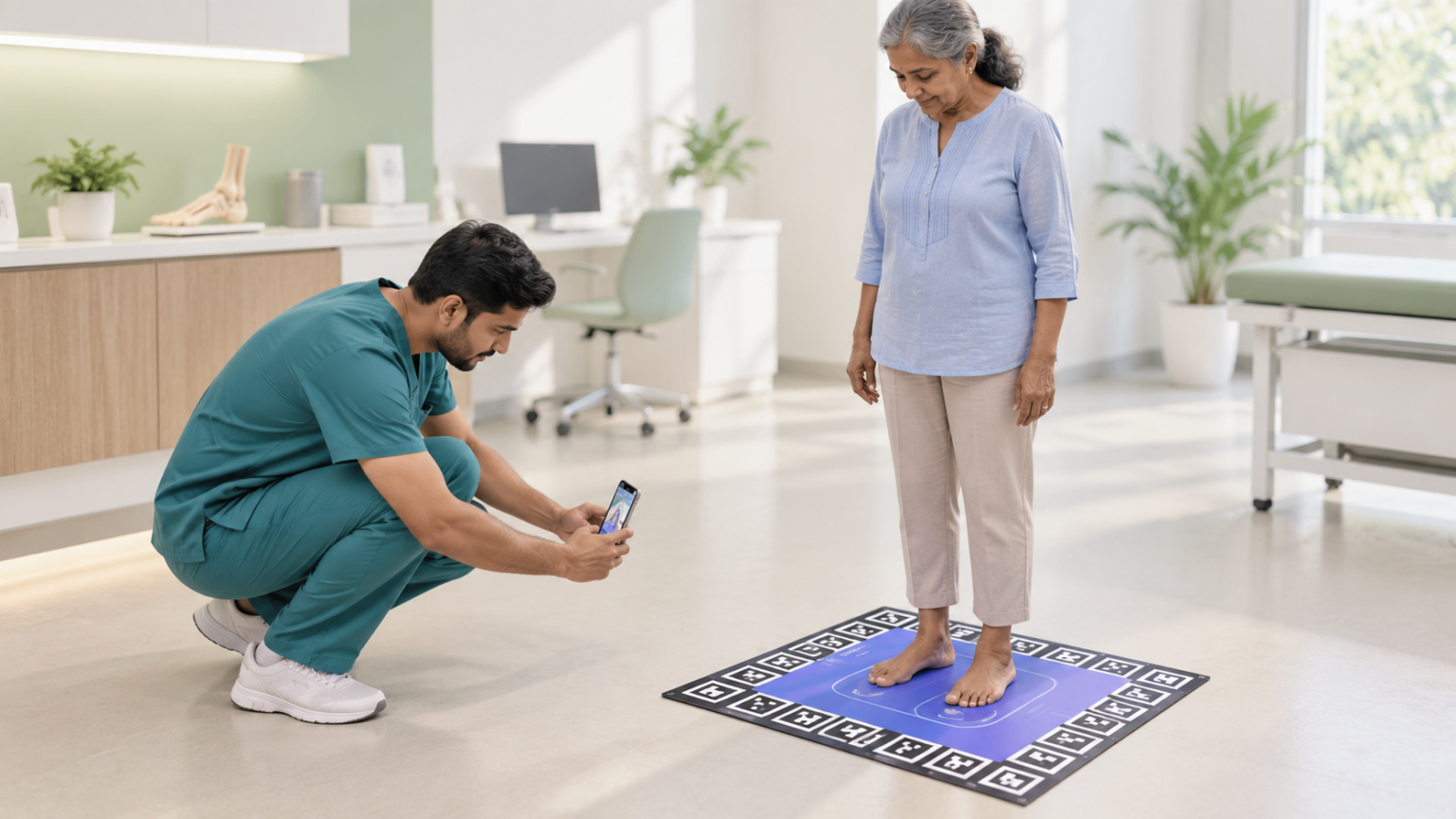
Foot scanning is a method of capturing the shape and dimensions of the foot and converting them into measurable information. Depending on the technology, it may generate a two-dimensional image or a three-dimensional (3D) surface model. Plantar-pressure systems serve a different purpose by assessing how load is distributed beneath the foot.
A digital foot model can provide measurements such as foot length, forefoot width, heel width, instep dimensions and selected arch-related parameters, depending on the scanning system. These measurements can help clinicians and footwear professionals understand individual fitting requirements and plan suitable footwear or insole modifications. The reliability of the information depends on the capture method, positioning, software and validation of the measurements.
2. The Growing Need for Individualised Foot Assessment
Foot morphology varies considerably between individuals. Differences in foot dimensions, arch contour, toe alignment, deformity and left-right symmetry can influence footwear fit and the space or support required during walking. Standard shoe sizes may not adequately represent these variations, particularly in people who require therapeutic footwear.
Medical footwear must often address more than shoe size. It may need to accommodate deformities, provide additional depth, protect vulnerable areas or incorporate a customised insole. Digital foot measurements can provide anatomical information for these decisions, while clinical examination and other relevant assessments establish the patient’s therapeutic requirements.
3. Clinical Relevance of Digital Foot Assessment
- Diabetes-related foot risk: People with diabetes may develop peripheral neuropathy, foot deformities and an increased risk of ulceration. Foot scanning can document foot shape and dimensions to support accommodative footwear and customised insole design. However, ulcer prevention requires comprehensive foot-risk assessment and appropriate pressure-relieving footwear. A shape scan alone cannot establish ulcer risk or confirm adequate pressure relief
- Foot deformities and structural variations: Conditions such as hallux valgus, hammertoes, flat feet and high arches can create specific footwear-fitting requirements. Digital measurements may help identify variations in width, toe shape and arch contour, supporting footwear designs that accommodate individual anatomy. The prescription should also consider symptoms, deformity severity and clinical findings.
- Musculoskeletal and specialised footwear needs: In selected patients with foot pain, altered biomechanics or post-surgical changes, foot scanning may help document anatomical variations and inform footwear or orthotic design. It contributes to the assessment of foot shape but does not independently diagnose the cause of pain or determine the appropriate treatment.
4. From Conventional Measurement to Digital Capture
Traditional foot assessment uses methods such as rulers, callipers, footprints, foam impressions and plaster casts. These techniques remain useful in clinical practice and orthotic fabrication. Manual measurements may record only selected dimensions, while physical impressions require additional handling and conversion into a design suitable for manufacture.
Dedicated 3D scanners capture the external surface of the foot and generate a digital model for measurement and computer-aided design. Research comparing 3D scanning with conventional methods has reported favourable precision for several foot dimensions, although performance varies by measurement and technique. Digital scanning should therefore be validated for the specific parameters and application for which it is intended.
5. Mobile App-Based Foot Scanning
Mobile app-based foot scanning combines a smartphone or compatible imaging device with software that processes foot images or depth data into measurements or a 3D model. Depending on the technology, the application may use multiple photographs, video, depth-sensing hardware or a combination of capture methods. This approach offers the potential to make digital foot measurement more accessible without requiring every assessment to take place at a dedicated scanning station.
For clinicians, a mobile workflow may support digital documentation and sharing of foot measurements. For footwear professionals, it may provide a digital reference for individualised fitting and insole design. These are potential workflow benefits rather than guaranteed clinical outcomes. Their achievement depends on measurement accuracy, capture quality and compatibility with the intended footwear-design process.
6. From Mobile Capture to a 3D Foot Model
- Image or depth-data capture: The user follows the application’s instructions to capture the foot from specified viewpoints. Image-based systems require sufficient overlapping views, while depth-enabled systems use distance information from compatible sensors.
- Foot detection and processing: Computer-vision algorithms identify the foot within the captured data and locate relevant anatomical landmarks. Blurred images, incomplete views, movement or unsuitable positioning can compromise the result.
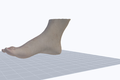
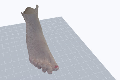
- Three-dimensional reconstruction: In image-based systems, software identifies corresponding features across multiple images and estimates their spatial positions to reconstruct a 3D point cloud or surface. Depth-based systems use measured distances to generate spatial points, which are subsequently aligned and processed into a model.
- Measurement extraction: Software calculates supported dimensions, such as foot length and width, using defined landmarks and measurement rules. A reliable scale reference and validated algorithms are essential for obtaining meaningful measurements.
- Digital output for design: The resulting model and measurements can be reviewed and transferred into compatible design software, where they may inform footwear fit, accommodation and insole geometry.
7. Evidence for Smartphone-Based Foot Measurement
Studies investigating smartphone-based foot measurement have reported promising results, demonstrating the potential of computer-vision technology to estimate foot dimensions such as length, width, arch height and instep girth from images and 3D scans. However, some evaluations have been conducted using a limited number of standardised foot models, and their findings may not fully reflect performance in real-world clinical settings.
Other research evaluating mobile applications for foot measurement has identified limitations in meeting the measurement requirements for custom-footwear manufacture. These findings highlight the need for further validation of measurement accuracy, repeatability and suitability for clinical and manufacturing applications before mobile scanning outputs are relied upon for individualised footwear design.
8. Translating Foot Measurements into Medical Footwear
A 3D foot model provides a digital representation of the foot’s external geometry. It can help document individual dimensions, asymmetry and contour variations that are relevant to footwear fitting. When compatible with the design workflow, the model may serve as an input for customised footwear or insole development.
The clinical prescription must combine digital measurements with patient assessment. For example, a person with diabetes and a history of plantar ulceration may require footwear that accommodates deformity and relieves pressure at high-risk locations. The scan can support anatomical fitting, while therapeutic requirements must be established through appropriate clinical assessment and pressure evaluation when indicated.
9. Research on Customised Footwear and Clinical Outcomes
Research highlights several potential benefits of integrating individual foot measurements, pressure-related information and clinical assessment into therapeutic footwear design.
Improved plantar-pressure distribution: Data-driven therapeutic footwear concepts have demonstrated reductions of 17-53% in peak pressure beneath the metatarsal heads compared with a non-therapeutic shoe, highlighting the potential of pressure-informed footwear design.
Footwear tailored to individual needs: Incorporating foot morphology, clinical risk and pressure-related information can help guide footwear design towards the specific requirements of people with diabetes and peripheral neuropathy.
Potential to support ulcer prevention strategies: Custom-made footwear designed to improve pressure relief may help reduce the risk of diabetic foot ulcer recurrence, particularly when patients wear the prescribed footwear consistently. A multicentre randomised trial found no significant reduction in overall ulcer recurrence in the intention-to-treat analysis, while a benefit was observed among participants with high footwear adherence.
A foundation for personalised footwear planning: Digital foot measurements can contribute to understanding foot dimensions and shape, supporting decisions about footwear fit and customised insoles. However, clinical assessment and appropriate pressure-relief evaluation remain essential to determine whether the final footwear meets the patient’s needs.
Foot scanning can contribute to the personalisation process, but improved clinical outcomes depend on the complete footwear intervention-including design, pressure redistribution, clinical suitability and patient adherence.
10. Foot Scan: Connecting Digital Measurement with Footwear Planning
Mobile Foot Scan has the potential to connect foot measurement, clinical review and personalised footwear planning within a digital workflow. By capturing foot geometry and processing it through dedicated software, such a system may help document individual dimensions and provide a digital reference for compatible footwear-design processes.
For clinicians, its potential value lies in providing anatomical information to support fitting decisions. For footwear professionals, it may offer a digital starting point for individualised footwear and insole design. For patients, it could simplify access to measurement-based footwear planning where appropriate clinical oversight and infrastructure are available.
The objective is to bridge the gap between measuring the foot and translating those measurements into a practical, individualised footwear solution. Clinical expertise remains essential in determining the appropriate footwear and therapeutic requirements.
11. Ensuring Clinically Meaningful Measurements
A clinically useful mobile foot-scanning workflow requires more than a realistic-looking 3D model. Measurement accuracy and repeatability should be validated against an appropriate reference method. Capture conditions, foot positioning, model completeness and the intended use of each measurement should be clearly defined.
The system’s claims should also remain within its validated capabilities. A shape scan should not be presented as measuring plantar pressure, circulation or sensation unless the system includes separate validated methods for those functions. For medical footwear, the most useful digital model is one that provides reliable measurements relevant to the clinical and manufacturing decisions being made.
BIBLIOGRAPHY
1. Bin Rafiq R, et al. Opti Fit: Computer-Vision-Based Smartphone Application to Measure the Foot from Images and 3D Scans. Sensors. 2022;22:9554.
2. Telfer S, Woodburn J. The use of 3D surface scanning for the measurement and assessment of the human foot. J Foot Ankle Res. 2010;3:19.
3. Gosa F, et al. Variable Lower Limb Alignment of Clinical Measures with Digital Photographs and the Footscan Pressure System. J Sport Rehabil. 2021;30(3):437–444.
4. Denckers F, et al. Foot shape assessment techniques for orthotic and footwear applications: a methodological literature review. Front Bioeng Biotechnol. 2024;12:1416499.
5. Jones PJ, et al. Foot Measurements from Three-Dimensional Scans: Elinvision 3DST Reliability. J Foot Ankle Res. 2025;18:e70070.
6. Lee YC, Lin G, Wang MJJ. Comparing 3D foot scanning with conventional measurement methods. J Foot Ankle Res. 2014;7:44.
7. Bus SA, et al. Effect of custom-made footwear on foot ulcer recurrence in diabetes: a multicentre randomized controlled trial. Diabetes Care. 2013;36:4109–4116.
8. Wafering JBJ, et al. Optimizing footwear for the diabetic foot: data-driven custom-made footwear concepts and their effect on pressure relief. PLOS ONE. 2020;15:e0224010.
9. Bus SA, et al. State of the art design protocol for custom-made footwear for people with diabetes and peripheral neuropathy. Diabetes Metab Res Rev. 2020;36(Suppl 1):e3237.
10. Kabir MA, et al. Mobile Apps for Foot Measurement in Pedorthic Practice: Scoping Review. JMIR Mhealth Uhealth. 2021;9(3):e24202.